Vaccine Aluminum Travels Into The Brain
http://vaccinepapers.org/al-adjuvant-nanoparticles-can-travel-brain/
FEB 10, 2015 |
“Parents can be reassured that the trace quantities
of aluminum in vaccines can’t possibly do harm.“
-Dr Paul Offit: Vaccine promoter, vaccine patent licensor, and self-appointed
autism pundit, 2015
Click for list of papers in this post.
Aluminum adjuvant nanoparticles (AANs) are transported through the body differently than ingested aluminum.
Most vaccines contain aluminum adjuvant, an ingredient necessary for stimulating a strong immune response and immunity. The aluminum is in the form of Al hydroxide and/or Al phosphate nanoparticles.
Aluminum has been used in vaccines since the 1920s. Despite this long history, aluminum adjuvant was not studied much beyond its effect of making vaccines more effective. The safety of injected Al adjuvant was assumed, largely because aluminum is a normal (if unhealthy) component of many foods. Its one of the most common elements of the Earths crust. Its everywhere.
So consideration of Al adjuvant safety was entirely based upon studies of ingested aluminum. Ingested aluminum has a low absorption (about 0.3%), and when this low absorption is taken into account, there is good reason to expect vaccines to create aluminum toxicity. But that is not the subject of the present commentary. Commentary about the total amount of aluminum in vaccines can be found here: http://vaccinepapers.org/danger-aluminum-vaccines/
The present commentary is concerned with the nanoparticulate form of aluminum in vaccines, and the evidence that aluminum adjuvant nanoparticles (AANs) can be far more toxic than ingested aluminum from food.
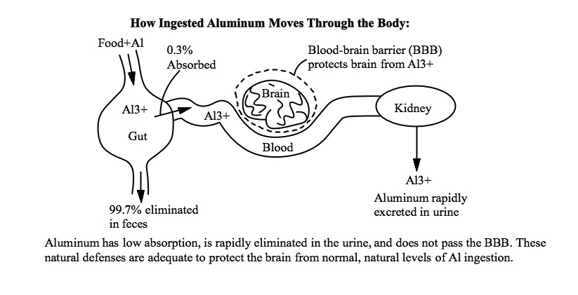
Ingested Aluminum
Ingested aluminum enters the blood from the gut. In the blood, ingested
aluminum is in a water-soluble ionic form, typically Al3+ or an aluminum
complex*. This aluminum is separated into individual atoms, like ordinary salt
dissolved in water. Ionic aluminum is toxic, but it is blocked from entering the
brain by the blood-brain barrier (BBB), and it is rapidly filtered from the
blood by the kidneys. Unless large amounts are consumed it does not cause a
problem.
Below is a diagram illustrating how ingested aluminum moves through the body. The body’s natural defenses are adequate for preventing harm from normal, natural amounts of ingested aluminum.
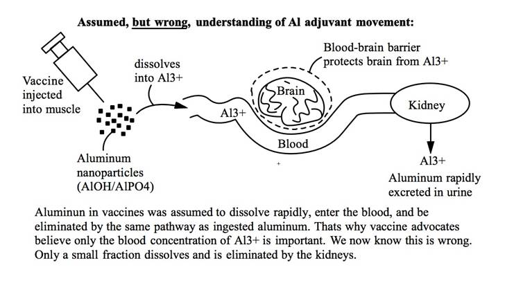
Based on on this understanding of ingested aluminum, it was long assumed that AAN follows a similar pathway out of the body. AANs cannot be filtered by the kidneys (they are too large). But it was assumed that the AANs would dissolve in body fluids, and the resulting Al3+ ions (or other Al ions like AlOH4-), would be filtered out by the kidneys, just like ingested aluminum. However, this simple model is wrong.
Below is a diagram illustrating this wrong understanding of how AANs travels through the body.
This model is wrong because what actually happens is that a type of white blood cell called a macrophage (MF) engulfs or “eats” (process is called “phagocytosis”) the AANs before they can dissolve. Eating foreign material is normal behavior for MFs. When MFs detect bacteria or other pathogens, the MFs engulf the pathogens, and digest them with enzymes. They then tell other immune system cells about the pathogen and how to detect it.
The problem with AANs is that they are not digested by the MF enzymes. And the AANs, once inside the MF, dissolve much more slowly. The AANs persist for a long time and cause the MFs to slowly leak aluminum. MFs that consume the AANs become highly contaminated with aluminum, and spread this aluminum around wherever they go. And they go everywhere in the body.
The MFs are able to travel across the blood brain barrier (BBB). The MFs, once loaded with AANs, act like a Trojan Horse and carry the AANs into the brain. This is very harmful, because the brain is very sensitive to aluminum.
Below is a diagram of a more correct model of how AANs travels around the body.
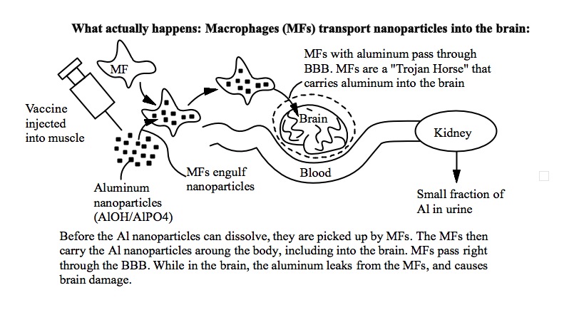
Once inside the brain, the aluminum damages brain cells. The damaged brain cells trigger inflammation which attracts more MFs, some of which are loaded with still more aluminum. The result is a vicious cycle.
The Scientific Evidence
The scientific evidence for this “Trojan Horse” mechanism is quite simple,
unequivocal and overwhelming.
First, there is the Flarend study, which shows that even after a month, only about 6% (of Al hydroxide) or 22% (of Al phosphate) are eliminated. Most aluminum adjuvant is retained in the body 1 month after injection (94% of Al hydroxide is retained!). The Flarend study also shows that the aluminum spreads to numerous organs, including the brain. The Flarend study did not determine the form of the retained aluminum, however (dissolved Al ions, or AANs). Flarend full paper: In vivo absorption of aluminium-containing vaccine adjuvants using 26Al
The Movsas study (published in 2013) used human infants and obtained similar results. Movsas looked for aluminum in urine and blood before and after routine vaccination with 1200mcg aluminum at the 2-month date. No change in urine or blood levels was observed. Movsas states:
“No significant change in levels of urinary or serum aluminum were seen after vaccination.“
Of course, these results contradict the claims by vaccine advocates that aluminum adjuvant dissolves and is removed by the kidneys. Movsas full paper: Effect of Routine Vaccination on Aluminum and Essential Element Levels in Preterm Infants
Several studies show, with certainty, that MFs engulf AANs. In several studies, the AANs have been stained and photographed inside the MFs, and identified using several different methods. This is not surprising because it is well known that MFs will engulf nanoparticles just from being grown in a solution containing nanoparticles. The composition of the nanoparticles does not seem to matter.
This paper describes an experiment proving that AANs are engulfed by human MFs, when grown in culture. See the photographs for yourself. Full paper: Unequivocal identification of intracellular aluminium adjuvant in a monocytic THP-1 cell line (Note: a macrophage (MF) is essentially the same as a “monocyte”. THP-1 is a specific macrophage culture used for research).
Another study shows that AANs are present inside MFs at the location of intramuscular vaccine injections. Human tissue samples were analyzed using X-rays, which identified high levels of aluminum inside the MFs. Full paper: Macrophagic myofasciitis lesions assess long-term persistence of vaccine-derived aluminum hydroxide in muscle.
In an impressive study in mice, AANs and other
nanoparticles (e.g. latex) were injected intramuscularly into mice. AANs were
detected in the brain and spleen, up to one year later. These results
contradict the long-assumed “100% of adjuvant dissolves into the blood”
belief preferred by vaccine advocates. Full paper: Slow
CCL2-dependent translocation of biopersistent particles from muscle to brain
(Note: CCL2 is an immune system signal that attracts MFs; it increases the
transport of the nanoparticles. The effect of CCL2 is further evidence that it’s
the MFs doing the transporting. CCL2 is described here: http://en.wikipedia.org/wiki/CCL2).
CCL2 is also known as “MCP-1” (macrophage chemo-attractant protein, a very
descriptive name since MCP-1 attracts macrophages). Below is a page from this
paper showing that AANs were detected in the brain and spleen.
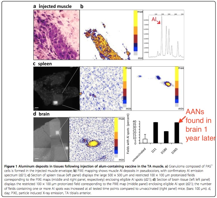
Images of AANs in brain and spleen. These particles traveled into the brain
from the distant site of intramuscular injection. AANs are not rapidly dissolved
and excreted, as vaccine advocates claim. D21, D180, D365 indicate time (in
days) between AAN injection, and detection in the brain.
Studies show that CCL2/MCP-1 production is stimulated by immune activation and
vaccines. So in the case of vaccines, AANs are injected at the same time the
immune system is producing CCL2/MCP-1. Hence, a vaccine provides all conditions
necessary for AAN transport into the brain. See our article on immune
activation here: http://vaccinepapers.org/part-1-immune-activation-autism/
Finally, the Trojan Horse mechanism of MF-transport of nanoparticles has been studied for applications in getting drugs into the brain (through the BBB). Studies have been done proving that nanoparticles, serotonin, or HIV drugs can be transported through the BBB using macrophages. In one specific study, the Trojan Horse mechanism was used to transport nanoparticles into a brain tumor (as a proof-of-principle experiment with the ultimate goal of transporting cancer drugs into the brain). In this study, human MFs engulfed nanoparticles (made of silica-gold). The particle-loaded MFs were then injected into mice (into the tail) and the particle-loaded MFs were detected in the brain tumor 24 hours later. Full paper: Delivery of nanoparticles to brain metastases of breast cancer using a cellular Trojan Horse .
QUOTES: (Choi et al., Delivery of Nanoparticles to Brain
Using Cellular Trojan Horse):
“More than two decades ago, Fidler and colleagues provided evidence that
macrophages of blood monocyte origin can infiltrate experimental brain
metastases while the blood–brain barrier is intact (Schackert et al. 1988). Even
earlier, Morantz and colleagues had quantified the content of macrophages in
clinical specimens (Morantz et al. 1979).”
AND
“The use of monocyte/macrophages as delivery vehicles to the CNS has been
investigated in situations other than malignancy. Afergan et al. demonstrated
the delivery of serotonin to the brain by monocytes, which had phagocytosed
nano-liposomes containing this otherwise brain impermeant drug (Afergan et
al. 2008). Dou and colleagues utilized bone marrow derived macrophages as
carriers of and depots for antiretroviral drugs to treat and attenuate the
symptoms of HIV-associated neurocognitive disorder (Dou et al. 2009). Therefore,
we hypothesized that nanoparticle-laden monocytes/macrophages would home in to
intracranial meta- static deposits by crossing the blood–brain barrier following
injection into the systemic circulation.”
Every step has been proven: MFs engulfing of Al nanoparticles, MFs crossing the BBB and MFs carrying nanoparticles into the brain has been experimentally demonstrated and proven multiple times. And the entire process has been demonstrated with AANs in mice. AANs from an intramuscular injection were “photographed” in the brain. These facts contradict the simplistic view (preferred by vaccine advocates) of aluminum toxicity based only on the concentration of dissolved aluminum ions in the blood. The story is far more complicated and worrisome than that.The toxicity of aluminum adjuvant depends critically on the transport of Al nanoparticles by MFs, through the blood-brain barrier.
Elevated MCP-1/CCL2 in Autistic Brain
Further, MCP-1/CCL2 is elevated in the autistic brain and spinal fluid. This was
one of the most significant findings of the Vargas study: Neuroglial
Activation and Neuroinflammation in the Brain of Patients with Autism.
Vargas stated:
“MCP-1 and TGF-B1 are the most prominent cytokines in
the brain of autistic patients.”
AND
“MCP-1, a chemokine involved in innate immune reactions and important mediator
for monocyte and T-cell activation and trafficking into areas of tissue injury,
appeared to be one of the most relevant proteins found in cytokine protein array
studies because it was significantly elevated in both brain tissues and
cerebro-spinal fluid.“
This of course means that in autism, injected AANs will be transported by MFs into the brain from a distant intramuscular injection site. MCP-1 attracts macrophages, and the macrophages are loaded with aluminum.
UPDATE: Nov 2015
A new study of Al adjuvant injections in mice has revealed even more complexity to the issue of Al adjuvant transport. As expected, it showed that Al adjuvant transport depends on MCP-1; mice that produce more MCP-1 (due to genetics) suffer greater transport into the brain.
Surprisingly, it also showed:
1) Transport depends on injection location. Subcutaneous injection
(i.e. under the skin) is necessary for brain transport, at least for the dosages
used. Intramuscular injection does not produce brain transport. This may be due
to the presence of more-mobile white blood cells (dendritic cells) in the skin
compared to muscle.
2) Transport depends inversely on dosage. A dosage of 200mcg/kg
resulted in brain transport (and behavioral changes) and dosage of 400mcg/kg did
not result in brain transport (and showed no behavioral changes). This may be
due to the high dosage impairing macrophage mobility. For example, higher local
inflammation at the injection site may cause reduced macrophage mobility.
There may also be an interaction between injection location and dosage. The dosage range that causes transport may be different for different tissues.
These phenomena may explain why the prior Al adjuvant injection experiments by the Shaw Laboratory (using 100, 300 and 550mcg/kg in divided doses) showed such strong adverse effects. Though aluminum adjuvant is harmful, in human infants the incidence of harmful effects is likely lower than what was observed in the prior mouse experiments. This has been a criticism made by vaccine advocates. Specifically, vaccine advocates have asserted that the reported effects were implausibly severe and therefore there must be something wrong with the prior studies by the Shaw Laboratory at UBC. This new study may explain why adverse effects in human infants are less frequent than what was observed in the Shaw Laboratory.
The authors state:
“In previously published studies, motor and behavioral
impairments were observed following sc (behind the neck) Alhydrogel® injection
to CD1 mice with doses of 100 and 300 μg Al/kg [17,41]. These effects were
associated with Al deposits in the central nervous system (spinal cord) assessed
by Morin stain. To examine if the route of exposure may represent an important
factor for alum toxicity, a nested study was conducted herein, showing that alum
particles may penetrate the brain at D45 after the sc (and not im) injection,
performed at the dose of 200 μg Al/kg (and not at the dose of 400 μg Al/kg). A
higher rate of brain translocation after sc injection may be explained by a much
higher density of dendritic cells with high migrating properties, in the skin
compared to the muscle. The fact that half dose resulted in brain
translocation, which was not observed at higher dose, is reminiscent of the
non-monotonic dose/response curves previously observed with environmental
toxins, including particulate compounds [67]. In another study, we similarly
observed neurobehavioral changes at 200 but not 400 μg Al/kg (Crépeaux et
al., manuscript in preparation). The exact significance of such observations is
unknown, but one may speculate that huge quantities of alum injected in the
tissue may induce blockade of critical macrophage functions such as migration
and xeno/autophagic disposition of particles, as previously reported for
infectious particles [37].”
sc=subcutaneous
im=intramuscular
Although this study does show that the adverse effects of Al adjuvant are less frequent than observed by the earlier experiments, it is confirmation of the Shaw laboratory results. This study is further evidence that Al adjuvant can cause brain damage at dosages human infants receive from vaccines.
Clearly, there is much more to learn about the dangers of Al adjuvant. The risk of Al adjuvant depends on genetics, on dosage in a complicated way, and on which tissue receives the injection.
Persistence
Another key finding from this study is the extreme biopersistence of Al adjuvant
particles. The particles were observed in distant organs and tissues up to 270
days after injection, including in the brain, spleen and lymph nodes. This is
confirmation of prior experiments that also found high persistence. Al adjuvant
nanoparticles dissolve only very slowly, if at all, and travel extensively
around the body.
The authors state:
“The present study confirms that alum is extremely
biopersistent [29, 37] and that alum biopersistence can be observed in both the
injected muscle and distant organs, including dLNs and spleen. Regarding the
strong immunostimulatory effects of alum and the unrequired depot formation for
its adjuvant activity [36], long-term biopersistence of alum in lymphoid
organs is clearly undesirable, and may cast doubts on the exact level of
long-term safety of alum-adjuvanted vaccines [37].”
dLNs = draining lymph nodes
alum = aluminum adjuvant
Full Paper (Crepeaux et al): Highly delayed systemic translocation of aluminum-based adjuvant in CD1 mice following intramuscular injections
If you would like to read more about this hugely important issue, read the following review paper by Dr Romain Gherardi (the researcher who made the photos above, of AANs in the brain and spleen of vaccine-injected mice). Paper is here: Biopersistence and brain translocation of aluminum adjuvants of vaccines
___________________________________________________________
NOTES:
AANs: Aluminum adjuvant nanoparticles. Used in most
vaccines.
BBB: Blood brain barrier. Protects brain from aluminum in normal conditions.
MF: Macrophage (same thing as monocyte). A type of white blood cell. Can travel
through the BBB.
CNS: Central nervous system (brain + spinal cord).
CCL2/MCP-1: Macrophage chemoattractant protein. Immune system signaling
substance that attracts MFs. Causes MFs to transport aluminum into the brain and
around the body.
* Under physiologic conditions, some dissolved aluminum will not be in the Al3+ form, but rather AlOH4-. For the sake of this discussion, this is irrelevant, so we will use Al3+, even though this is not really correct. But AlOH4- arguably contains Al3+ in the center.
Monocytes and macrophages are basically the same thing. From nature.com: “Macrophages (and their precursors, monocytes) are the ‘big eaters’ of the immune system. These cells reside in every tissue of the body, albeit in different guises — such as microglia (brain), Kupffer cells (liver) and osteoclasts (bone) — where they engulf apoptotic cells and pathogens and produce immune effector molecules. Upon tissue damage or infection, monocytes are rapidly recruited to the tissue, where they differentiate into tissue macrophages. Macrophages are remarkably plastic and can change their functional phenotype depending on the environmental cues they receive. Through their ability to clear pathogens and instruct other immune cells, these cells have a central role in protecting the host but also contribute to the pathogenesis of inflammatory and degenerative diseases.”
Papers in this post:
Slow CCL2-dependent translocation of biopersistent particles from muscle to brain
Khan Z, Combadière C, Authier FJ, Itier V, Lux F, Exley
C, Mahrouf-Yorgov M, Decrouy X, Moretto P, Tillement O, Gherardi RK, Cadusseau
J. BMC medicine 2013 Apr 4;11:99.
PubMed Link
Delivery of nanoparticles to brain metastases of breast cancer using a cellular Trojan horse
Choi, Mi-Ran. Bardhan, Rizia. Stanton-Maxey, Katie J.
Badve, Sunil. Nakshatri, Harikrishna. Stantz, Keith M. Cao, Ning. Halas, Naomi
J. Clare, Susan E. Cancer nanotechnology 2012; 3(1-6):47-54
PubMed Link
Gherardi, R K. Coquet, M. Cherin, P. Belec, L. Moretto,
P. Dreyfus, P A. Pellissier, J F. Chariot, P. Authier, F J. Brain : a journal of
neurology 2001; 124(Pt 9):1821-31
PubMed Link
Unequivocal identification of intracellular aluminium adjuvant in a monocytic THP-1 cell line
Mold, Matthew. Eriksson, Håkan. Siesjö, Peter. Darabi,
Anna. Shardlow, Emma. Exley, Christopher. Scientific reports 2014; 4():6287
PubMed Link
Neuroglial Activation and Neuroinflammation in the Brain of Patients with Autism
Vargas, Diana L. Nascimbene, Caterina. Krishnan, Chitra.
Zimmerman, Andrew W. Pardo, Carlos A. Annals of neurology 2005; 57(1):67-81
PubMed Link
In vivo absorption of aluminium-containing vaccine adjuvants using 26Al
Flarend, R E. Hem, S L. White, J L. Elmore, D. Suckow, M
A. Rudy, A C. Dandashli, E A. Vaccine ; 15(12-13):1314-8
PubMed Link
Biopersistence and brain translocation of aluminum adjuvants of vaccines
Gherardi, Romain Kroum. Eidi, Housam. Crépeaux,
Guillemette. Authier, François Jerome. Cadusseau, Josette. Frontiers in
neurology 2015; 6():4
PubMed Link
Effect of Routine Vaccination on Aluminum and Essential Element Levels in Preterm Infants
Movsas, Tammy Z. Paneth, Nigel. Rumbeiha, Wilson.
Zyskowski, Justin. Gewolb, Ira H. JAMA pediatrics 2013; 167(9):870-2
PubMed Link